
Digital Screening Mammograph
Digital Screening Mammograph continues to be a major global health concern, affecting millions of women worldwide. Timely detection and early intervention are crucial in improving survival rates and treatment outcomes. In recent years, advancements in medical imaging technology have revolutionized breast cancer screening, with digital screening mammography emerging as a powerful tool in the early detection of breast abnormalities.
What IS Digital Screening Mammograph:
Digital Screening Mammograph This article explores the benefits and advancements of digital screening mammography, highlighting its potential to enhance breast cancer detection and improve patient outcomes.
The Evolution of Mammography:
Mammography, a specific type of X-ray imaging, has long been recognized as the gold standard for breast cancer screening. Traditional film-based mammography has been the primary diagnostic tool for decades. However, this technique had limitations such as lower image quality, longer processing times, and difficulties in transmitting and storing images. These shortcomings prompted the development of digital mammography.
Digital Screening Mammography Explained:
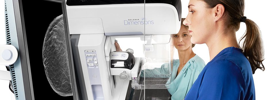
Digital screening mammography utilizes electronic detectors to capture and convert X-ray images into digital signals, which can then be viewed and manipulated on computer screens. The transition from analog to digital technology offers several significant advantages. Firstly, digital images can be instantly obtained, eliminating the need for film processing and reducing patient waiting times. Furthermore, digital images can be easily stored, retrieved, and shared electronically, streamlining communication between healthcare providers and enhancing the continuity of care.
Improved Image Quality and Interpretation:
One of the key benefits of digital screening mammography is the improved image quality it provides. Digital images offer higher resolution and improved contrast, allowing radiologists to detect smaller lesions and subtle abnormalities more effectively. The enhanced clarity and detail offered by digital mammography contribute to increased diagnostic accuracy, reducing the likelihood of false-positive or false-negative results.
Computer-Aided Detection (CAD) Systems:
Digital screening mammography has also paved the way for the integration of computer-aided detection (CAD) systems. CAD systems use advanced algorithms to analyze mammographic images and identify potential areas of concern. These systems act as a "second pair of eyes" for radiologists, highlighting regions that may require closer inspection. CAD systems have shown promising results in improving breast cancer detection rates and reducing interpretation errors, providing radiologists with valuable support in the diagnostic process.
3D Digital Breast Tomosynthesis:
In recent years, 3D digital breast tomosynthesis (DBT) has emerged as a significant advancement in digital mammography. DBT creates a series of thin cross-sectional images of the breast, allowing radiologists to examine breast tissue layer by layer. This technique overcomes the limitations of traditional 2D mammography, reducing overlapping structures and improving lesion visibility. DBT has demonstrated increased cancer detection rates and a reduction in false-positive recalls, enhancing both sensitivity and specificity in breast cancer screening.
Digital Mammography: An Opportunity for Personalized Medicine:
Digital screening mammography also holds the potential to facilitate personalized medicine approaches. With digital images readily available, radiologists can use computer-based tools to analyze breast density, an important risk factor for breast cancer. This information can be integrated into personalized risk assessment models, enabling tailored screening and surveillance strategies for individual patients. Furthermore, digital mammography allows for the integration of machine learning algorithms, which have the potential to improve the accuracy of risk prediction and aid in the early identification of high-risk individuals.
Conclusion:
Digital screening mammography has transformed the field of breast cancer screening, offering numerous advantages over traditional film-based techniques. The improved image quality, integration of CAD systems, and the emergence of 3D digital breast tomosynthesis have significantly enhanced the detection of breast abnormalities. Moreover, digital mammography provides opportunities for personalized medicine approaches, contributing to more targeted and effective breast cancer screening and management strategies. As technology continues to advance
Digital Screening Mammograph How Its Work
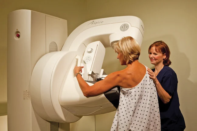
Digital screening mammography works by utilizing electronic detectors to capture X-ray images of the breast and convert them into digital signals. Here's a step-by-step breakdown of how the process works:
- Preparation: Before the mammogram, the patient is instructed to remove any jewelry or clothing that could interfere with the imaging process. The technologist then positions the patient and the breast on a dedicated mammography machine.
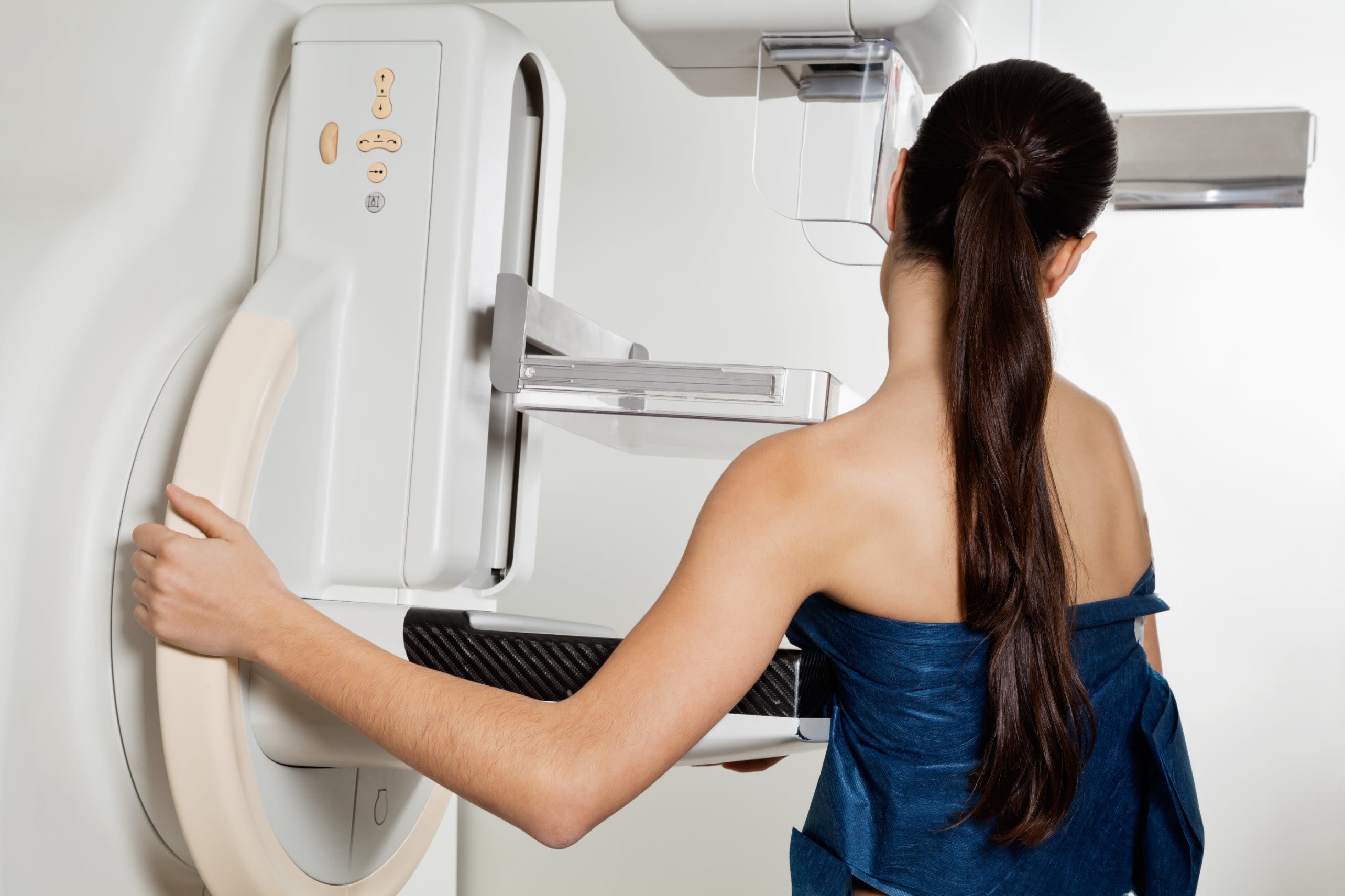
- Image Acquisition: The breast is compressed between two plates to spread out the tissue and obtain a clear image. Compression is necessary to reduce motion and improve image quality. The technologist positions the breast and adjusts the machine as needed to capture images of both the top-down (craniocaudal) and side-to-side (mediolateral oblique) views. Multiple images may be taken of each breast to ensure comprehensive coverage.
- Digital Conversion: Instead of using X-ray film, digital mammography uses electronic detectors that capture the X-ray energy passing through the breast tissue. These detectors convert the X-ray energy into digital signals.
- Image Processing: The digital signals are sent to a computer, where they are processed and reconstructed into high-resolution digital images. The images can be adjusted for brightness, contrast, and magnification to optimize visualization of breast tissue.
- Image Review: The radiologist reviews the digital images on a computer monitor. They examine the breast tissue for any signs of abnormalities, such as masses, calcifications, or architectural distortions. The enhanced clarity and detail of digital mammography images aid in the detection of subtle abnormalities.
- Computer-Aided Detection (CAD): In some cases, a computer-aided detection (CAD) system may be utilized. CAD systems use advanced algorithms to analyze the mammographic images and highlight areas that may require closer inspection. CAD acts as a "second pair of eyes" for radiologists, assisting in the detection of potential abnormalities.
- Follow-up and Diagnosis: If the radiologist identifies any suspicious findings, further diagnostic tests such as additional mammographic views, ultrasound, or biopsy may be recommended to determine if the abnormalities are cancerous or benign.
It's important to note that digital screening mammography is a safe procedure, and the level of radiation exposure is low. The benefits of early detection and accurate diagnosis outweigh the minimal risks associated with radiation exposure.
If you want to get amazing benefits by using this link
Final Words:
Overall, digital screening mammography offers significant advantages over traditional film-based mammography, including faster image acquisition, improved image quality, easier image storage and retrieval, and the potential for computer-aided detection systems to enhance detection rates. These advancements contribute to more effective breast cancer screening and early intervention, ultimately improving patient outcomes.